X-ray

Carrying out the radiographic examination

Indications for radiography

The difference between x-ray and ultrasound
X-ray
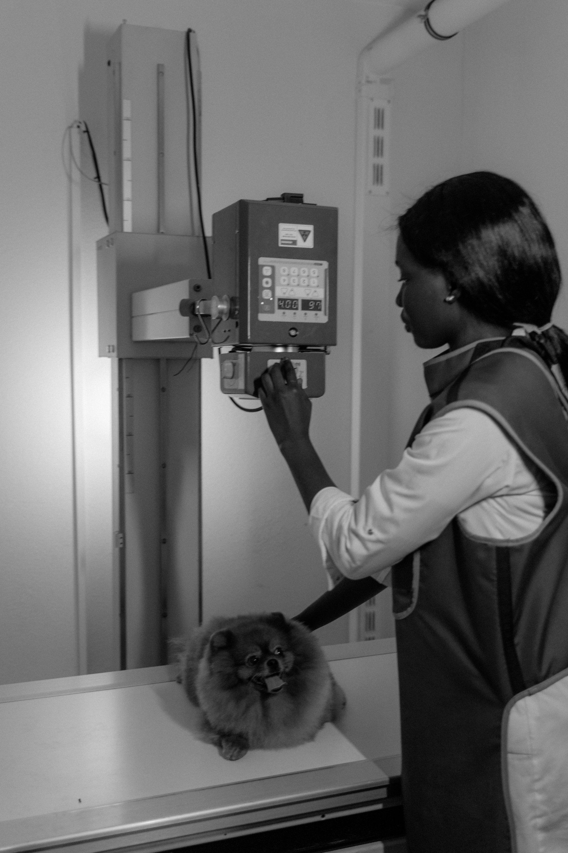
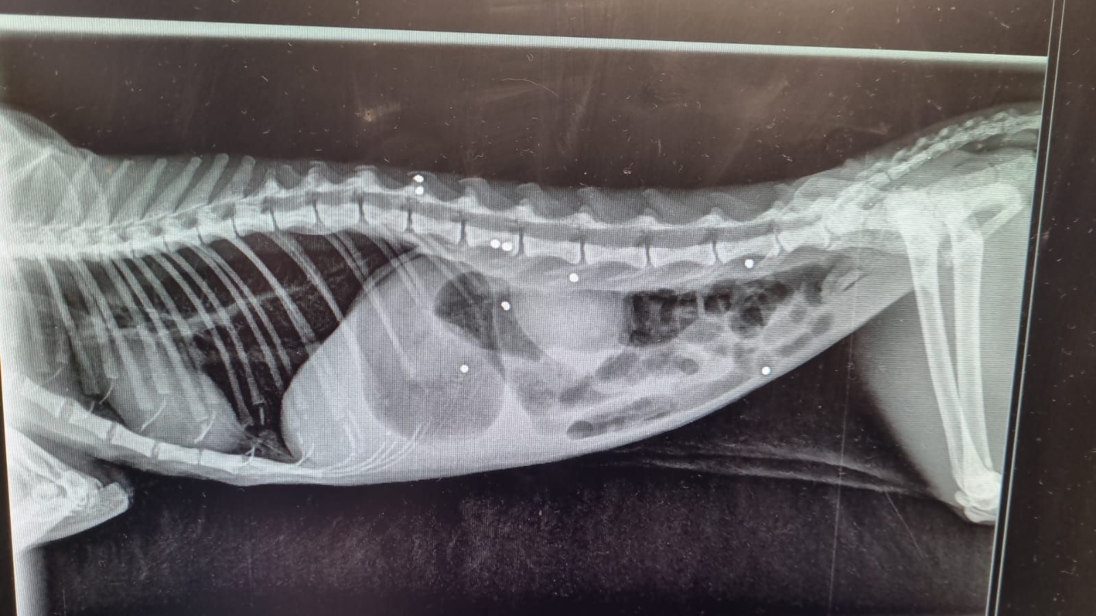
The X-ray machine emits X-rays, while the sensor receives them. The rays will then pass through the body and print an image of the latter on the sensor.
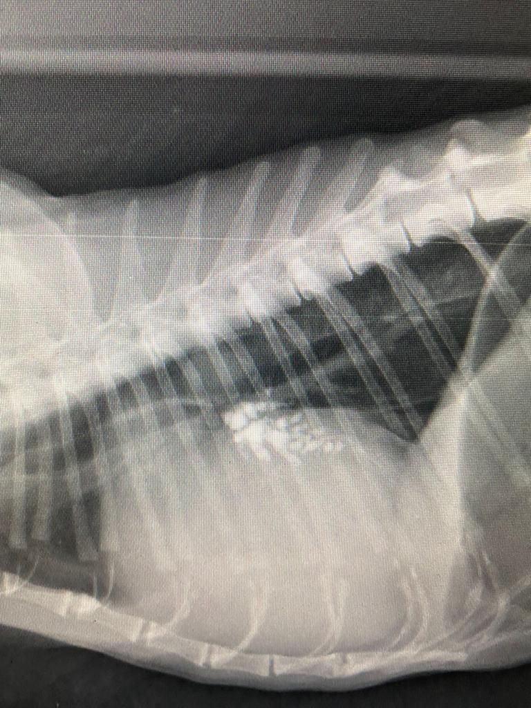
The image is fixed here, we can correctly observe the bone structures which is not the case with ultrasound but on the contrary we cannot see the movement of the organs.
Ultrasound
Ultrasound uses a probe that emits short pulses of ultrasound into the body and, within a fraction of a second, receives multiple echoes back from the part of the body being examined.
These echoes are converted into detailed images of internal organs and tissues, visible on the screen. The application of a gel amplifies the transmission of ultrasound and improves the quality of the images thus obtained.
The big difference between the two techniques is that ultrasound is a dynamic examination where we can see the movements of certain organs or blood flows, unlike x-rays which are a fixed image.


